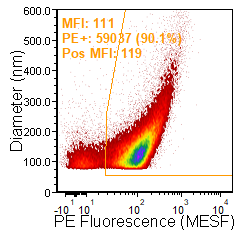

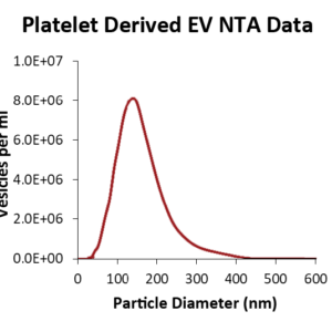
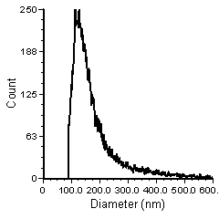
![Validation of Anti-CD9 vTag™ Antibody HI9a [PE] in vFC™ staining. (A) vFC™ count and size of platelet derived extracellular vesicles (EVs), a positive control sample for CD9 staining; (B) Anti-CD9 vTag™ staining of platelet derived EVs (120 second acquisition time, for more details see our vFC™ assay and protocol); (C) Anti-CD9 vTag™ staining of negative control samples (Lipo100™); (D) Positive (B) and negative (C) control data summarized and merged for comparison.](https://cdn-cellarcus.pressidium.com/wp-content/uploads/2019/04/CD9-vTag-EV-staining-data.jpg "CD9 vTag EV staining data")
vTAG™ ANTI-HUMAN CD9 ANTIBODY
NO WASH, QUANTITATIVE CD9 MEASUREMENT BY vFC™
$105.00 – $295.00
|
Conjugate(s): PE
Est. Limit of Detection: 30 Molecules per EV
Cargo analysis using vTag™ antibodies should be accompanied by a vesicle specific analysis method such as vFC™. vTag™ antibodies bundled with the vFC™ assay will be discounted 25%. |
Certificate of Analysis
|
||
 TDS TDS |
 MSDS MSDS |
||
Description
CD9 Immunostaining in vFC™?
vFC™ (#CBS4) is an assay that enables specific detection and characterization of individual extracellular vesicles such as exosomes and microvesicles on commercially available flow cytometers. This protocol makes use of vFC™ to provide a standardized means of analyzing individual vesicles to measure size, concentration, and CD9 expression simultaneously. Because vFC™ triggers on vesicles, pairing antibody staining with vFC™ enables a highly-reproducible, no-wash assay. In this protocol, vesicles will be stained using our vTag™ Anti-Human CD9 Antibody. See protocol 2 in the vFC™ kit handbook for additional considerations and full protocol details.
Procedure
- Dilute sample containing EVs to approximately 5×10⁶ extracellular vesicles per µl. To empirically determine vesicle concentration, see supporting protocols included in the vFC™ kit.
- Prepare a working concentration of vFluor™ Red (5 µl per well) according to kit instructions
- In duplicate, in the included 96 well plate, add 35 µl of dilution buffer, 5µl of vFluor™ Red, 5 µl of vTag™ anti-human CD9 antibody, and 5 µl of EV sample.
- Incubate in the dark for 1hr.
- Perform serial dilution of the samples as described in the assay handbook. Briefly, samples are to be diluted from between 1:100 to 1:1000 from staining concentration depending upon the flow cytometer being used.
- Analyze the diluted sample of labeled EVs on a qualified flow cytometer.
CD9 is a member of the tetraspanin family. In cells, tetraspanins are importnat to many biological functions including cell adhesion and motility as well as signaling and protein trafficking.
CD9 was first reported in EVs from dendritic cells (which were described as exosomes). It is abundant in the membrane of many cells and is present in larger membrane EVs. CD9 has broad tissue distribution and is one of several molecules previously thought to be useful as a pan exosome or extracellular vesicle marker. Of course, we now know that EVs from different sources exhibit different tetraspanin expression profiles which suggests that different tetraspanins will serve as markers for different vesicle subpopulations. For instance, in our own work profiling EV cargo in vesicles originating from distinct subsets of cells, we have found that CD9 is highly expressed in vesicles derived from various immune cell populations, certain carcinoma cell lines, and platelet derived EVs. However, CD9 expression is very low or absent in vesicles derived from red blood cells and the glioblastoma cell line U87MG (see the data by vesicle origin resource for more info).
-
vFC™ VESICLE FLOW CYTOMETRY EV ANALYSIS ASSAY
$450.00 – $2,100.00 Select options This product has multiple variants. The options may be chosen on the product page
FOR COUNTING AND SIZING VESICLES IN BIOFLUIDS, MEDIA, OR BUFFER -
HUMAN PLATELET DERIVED EXTRACELLULAR VESICLES
$275.00 – $1,300.00 Select options This product has multiple variants. The options may be chosen on the product page
Additional information
| Size | 20 Tests, 100 Tests |
|---|---|
| Conjugate | PE, PE-Cy7 |
Reviews
There are no reviews yet.